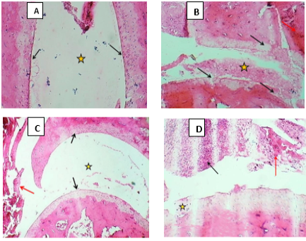
Microscopically examination of joint tissue in A-control negative: section of joint shows normal joint cavity (Asterisk) & normal articular surface (Black arrows). B- (G1) section of joint shows filled of joint cavity with necrotic tissue debris (Asterisk), degeneration of articular cartilage (Black arrows) & thickening of articular capsule (red arrow).C- (G2) section of joint that shows normal joint cavity with normal articular cartilage (Black arrows), normal outer fibrous joint capsule with normal inner synovial membrane (Red arrow). D- (G3) section of joint that shows moderate arthritis characterized by degeneration with necrosis of once of articular cartilage with marked infiltration of MNCs (Black arrow), irregular articular surface (Asterisk). H&E stain. 100x