
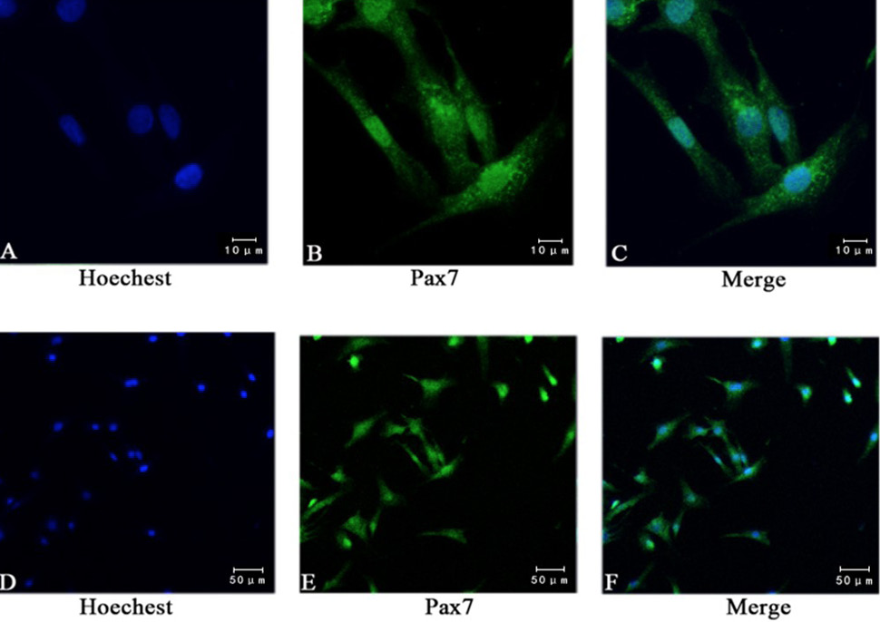
Fig. 2.
Immunofluorescence staining showed the expression of pax7. (A, D) Staining nuclei with Hoechst; (B, E) Using anti pax7 antibody immunofluorescence staining, 95% of the cells were positive; (C, F) Pax7 and Hoechst composite picture. (A, B, C bar = 10 μm; D, E, F bar = 50 μm).