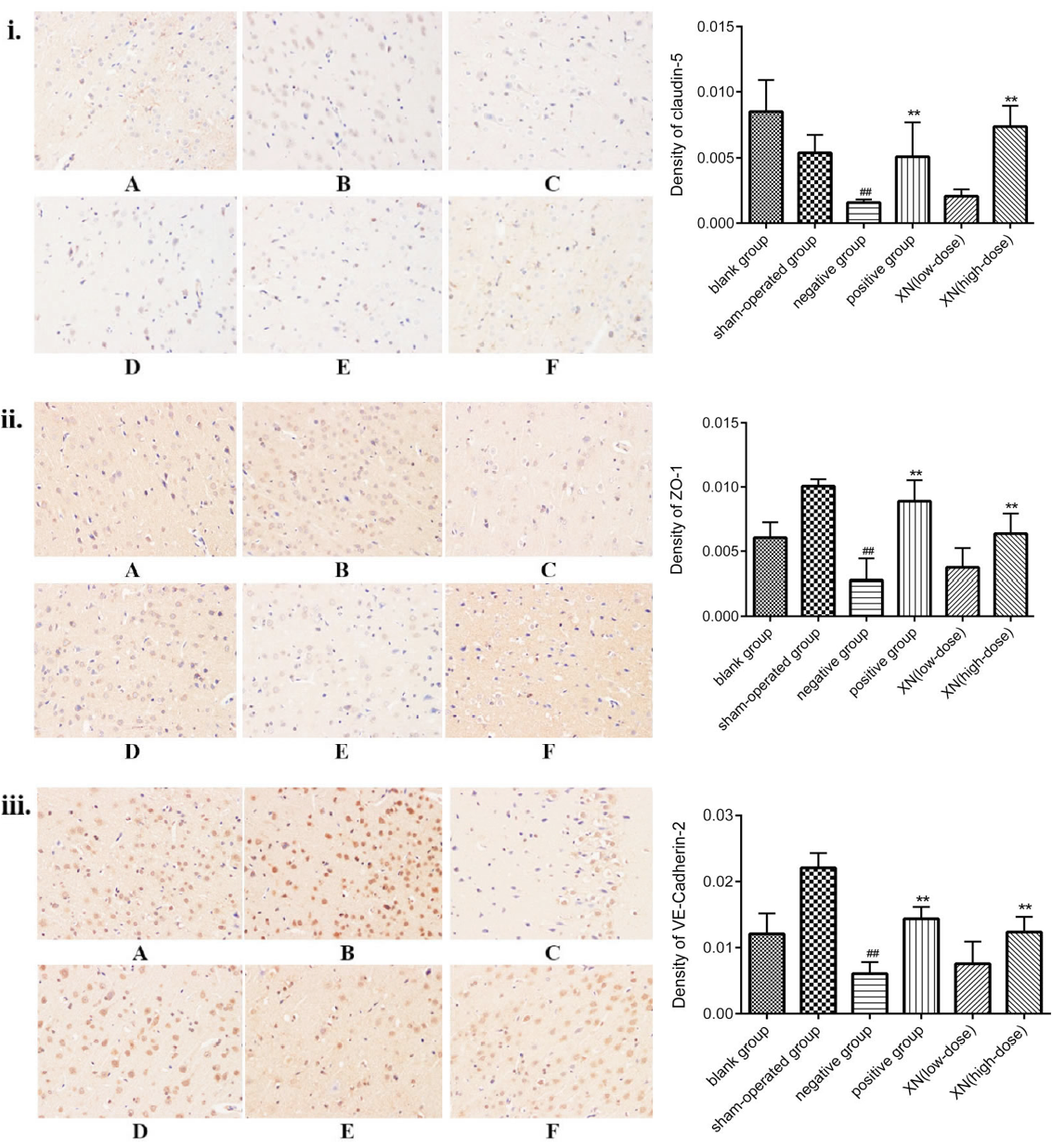
Expression alteration of claudin-5, ZO-1 and VE-cadherin protein by immunohistochemistry in ICH rat hippocampus tissue in blank group (A), sham-operated group (B), model group (C), AGNH group (D), and XN-treated (low/high doses, E/F) groups. All images were taken at 400× magnification. (i) expression of Claudin-5 protein in hippocampus of ICH rats; (ii) expression of ZO-1 protein in hippocampus of ICH rats; (iii) expression of VE-cadherin protein in hippocampus of ICH rats. The results are also expressed in a bar graph showing the density alteration of claudin-5, ZO-1 and VE-cadherin protein in hippocampal tissue. Data are represented as mean±SEM (#P<0.05, ##P<0.01, versus sham-operated group; *P<0.05, **P<0.01, versus model group); data analyzed from six independent experiments (n=6).
