
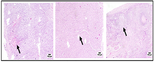
Figure 3
Histological evaluation of different stages of CL (early, mid and late stages). a) Photomicrograph of early stage of corpus hemorrhagicum, showing blood filled cavities (arrow) (H&E). b) Photomicrograph of mid stage of corpus luteum, showing complete luteinization of granulosa cells with existence of connective tissue septa (arrow) (H&E). c) Photomicrograph of late stage of corpus luteum, showing development of fibrotic masses (arrow) (H&E).