
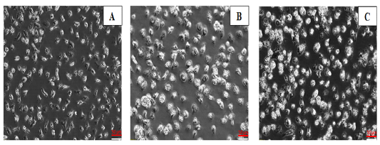
Figure 3
Morphologies of chBM-DCs for (A) non-infected control cells, (B) WT FWPV-infected cells, and (C) rFWPV-H5 infected cells, cultured in the presence of GM-CSF and IL-4, using a phase-contrast inverted microscope. Bar: 50 µm. Magnification: 200X.