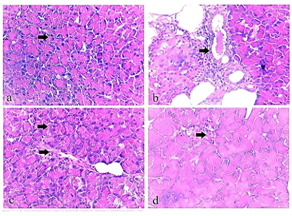
Figure 16
Photomicrograph of comparative figure of the normal group showed normal architecture of the pancreas (Fig. 16 a). The pancreas of group 2 showed severe congestion and thickening of blood vessels with inflammatory edoema (Fig. 16 b). The pancreas of group 3 showed haemorrhage with RBCs infiltration and mild congestion of blood vessels (Fig. 16 c). The pancreas of group 4 showed minimal vacuolation of pancreatic islets (Fig. 16 d).