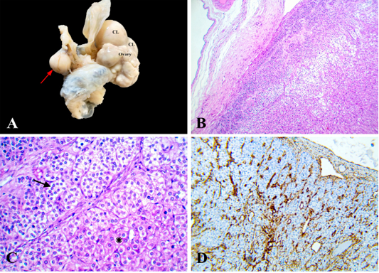
Figure 2
Interstitial cell tumor, ovary, camel. (A) Firm grayish white nodule (arrow) attached to mesovarium, CL; corpus luteum. (B) Encapsulated moderately cellular neoplasm, HE x 100. (C) Neoplastic cells arranged in solid sheets, cords (asterisk), and indistinct nests or acini (arrow) separated by fine fibrovascular stroma, HE x 400. (D) Neoplastic cells expressed cytoplasmic positive reaction for vimentin, x 200.