
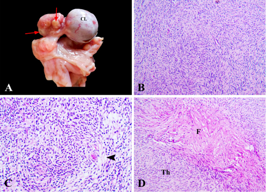
Figure 3
Thecoma, ovary, camel. (A) Small firm grayish white nodules (arrow), CL; corpus luteum. (B) Aggregates of streaming oval and spindle cells arranged in a diffuse pattern, HE x 200. (C) Neoplastic cells had pale vacuolated cytoplasm (arrowhead), HE x 400. (D) Fibrothecoma characterized by mixtures of fibroma (F) and thecoma (Th), HE x 200.