
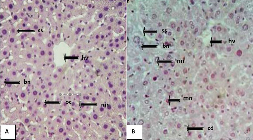
Figure 1:
Photomicrographs from representing sections of the liver of Cnt and ATZ treated groups (H and E staining at 400×). Cnt: (A) liver exhibited a well-organized structure of trabeculae radiating from hepatic veins (hv) having mononucleated (mn) and binucleated (bn) hepatocytes separated by regular sinusoidal spaces (ss). ATZ treated group (B): liver showed the disorganized structure of trabeculae arising from the hepatic vein (hv) with deformed shape mononucleated hepatocytes (mn), binucleated hepatocytes with swollen nuclei (bn), cellular degeneration (cd), dilated sinusoidal spaces (ss), hollow and necrotic nuclei (nn) were also observed in the treated liver.