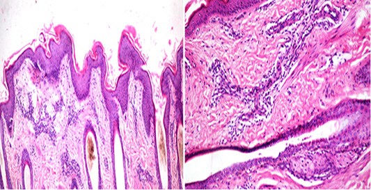
Figure 3
Histopathology of Bovine skin showing (a) Edema, separation, and moderate disorganization of collagen bundles in the dermis in addition to perivasculitis (Masson’s Trichrome stain X 40), (b) loss of spatial orientation of grains and dislodgement of poorly stained collagen bundles (Immunoperoxidase and Mayer’s Hematoxylin counterstain X 400).