
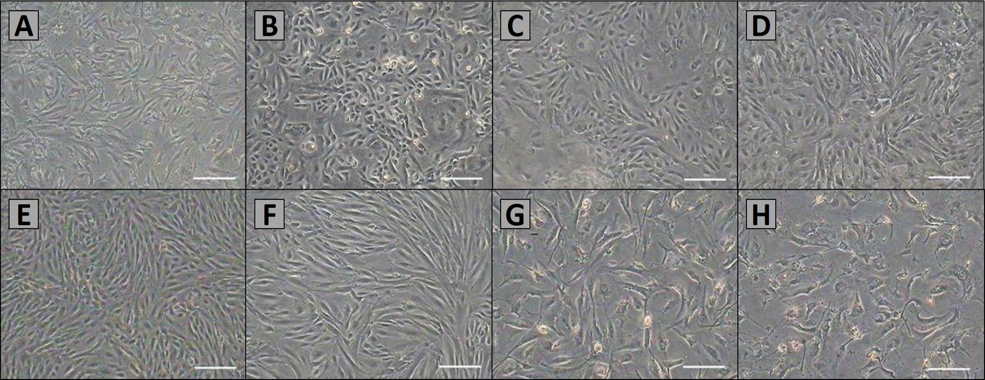
Fig. 1.
Morphology of primary and sub-cultured AMSCs. A, primary cells were cultured after 48 h. Most of cells began to adhere and stretch; B, AMSCs were cultured about 4 days, the cells grew to 70%–80% confluence; C, the cells of P1 were non-homogenous; D, after P5, AMSCs were purified, and there was apparently no difference in morphology among successive passages. AMSCs exhibited a fusiform pattern; E and F, morphology of P5, P10 and P15 AMSCs; G and H, the AMSCs of P24 and P25 displayed a typical senescence. Scale bar, 100μm.