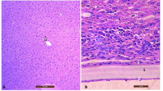
Figure 2
(A) Sections from the liver of the control uninfected mouse showed central vein (arrow) and hepatic cells(10X), Scale bar= 10µm. (B) Section of mice infected liver with hydatid cyst showing characteristics cyst layers laminated layer (L) and germinal layer (G) surrounded by fibrous tissue (F) with intense inflammatory cell infiltration (IC). (40X), Stained with H&E. Scale bar = 2µm.