
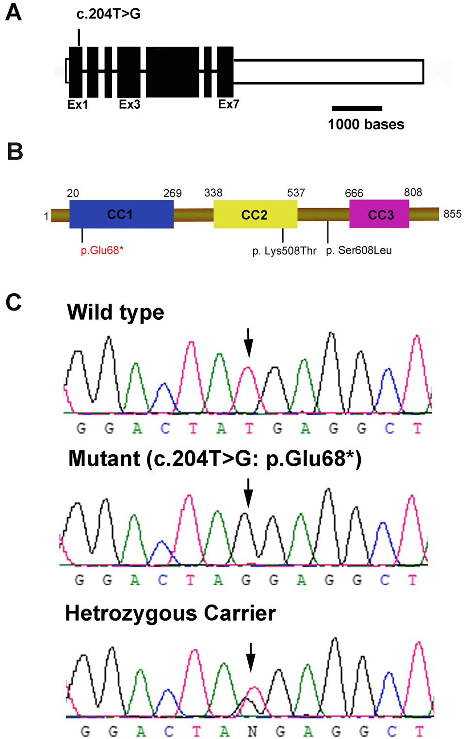
Fig. 3.
Mutation identification (A) BICD2 gene structure showing 7 exons. The mutation identified is indicated above in exon1, unfilled boxes at the start and end of the gene represents UTR regions. (B). BICD2 protein structure (855 amino acids). Domains are indicated by the specified color. N-terminal CC1 domain interacts with dynein-Dynactin complex, CC2 central domain interacts with kinesin protein and CC3 at C-terminal interacts with RAB6A, RNBAP2. Variants identified in HSP families so far are shown below, mutation identified in this study is in red, while text in black indicates variants identified earlier. (C). Chromatograms showing mutation in patient, heterozygous carrier along with wild type control.