
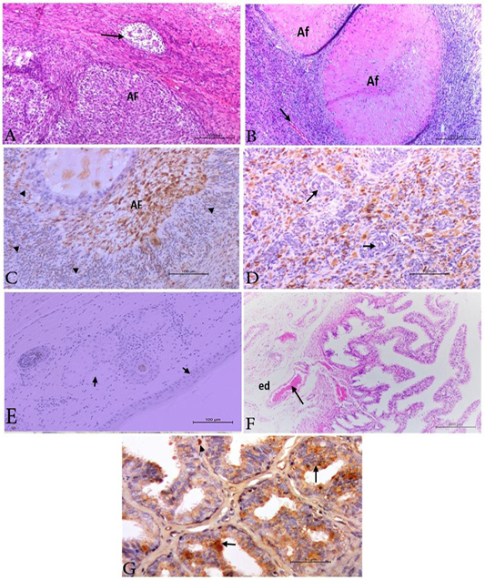
Photomicrograph from ovary, ear notch and uterine tube of acute infected animals. A) Ovary with collapsed atretic follicle (AF) and focal granulosa cell tumor (arrow) .H&E. Bar. 200µm. B) Ovary with collapsed atretic follicles (AF) and congested stromal blood vessels (arrow).H&E. Bar. 200µm. C) Ovary revealed positive BVDV antigen in the collapsed atretic follicle (AF) and in macrophages at the ovarian stroma. IHC. Bar.100µm. D) Ovary with positive macrophages at the ovarian stroma while the blood vessels appear clear negative (arrow). H&E. Bar.50 µm. E) Ear notch showing negative reaction for BVDV antigen (arrow). IHC. Bar.100 µm. F) Infundibulum showing marked folding of mucosa –submucosa layer and severe congestion of the vasculature (arrow). also edema (ed) at the tunica serosa and musculosa. H&E. Bar.200µm. G) Uterine tube showing positive BVDV antigen in the epithelium in focal manner (arrow) and in macrophages at the sub mucosa (arrow head). IHC. Bar.50µm.