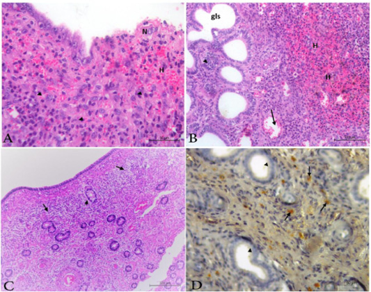
Figure 7
Photomicrograph from uterus of acute infected animals showing, A) Necrosis (N); sloughing of mucosa; diffuse infiltration of mononuclear cells (arrow head); congestion (arrow) and hemorrhages (H). H&E. Bar.50µm. B) Necrosis; hyperplasia of the endometrial gland (arrow head) associated with Cystically dilated glands (gls) beside congestion (arrow) and hemorrhages (H). H&E. Bar.100µm. C) Chronic endometritis presented by granulation tissue (arrow) and periglandular fibrosis at the endometrial stroma (arrow head).H&E. Bar.200µm. D) brown granules represent the positive BVDV antigen (arrow). All uterine glands are free from BVDV antigen (arrow head).H&E. Bar.50µm.