
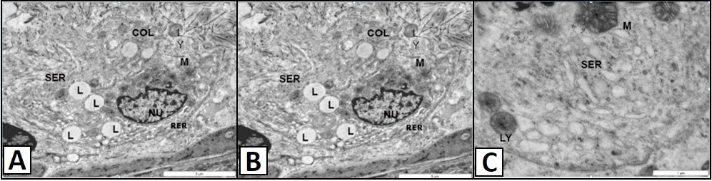
TEM micrograph showing of the Leydig cells of the vervet monkey, Chlorocebus aethiops. A, shows the polygonal nucleus (NU) has evenly distributed heterochromatin patches. The rough endoplasmic reticulum (RER) is situated at the basal region of the nucleus. The smooth endoplasmic reticulum (SER) is located at the apical region of the nucleus. The lipid droplet (L) and the lysosomes (LY) are found scattered in the cytoplasm together with collagen fibers (COL). B, show the distinct mitochondria of the Leydig cells. B, shows the smooth endoplasmic reticulum (SER) is located at the apical region of the nucleus. The lysosomes (LY) are found scattered in the cytoplasm. C, shows the tubular cristae (C) are clearly observed forming the mitochondria. The lipid droplets (L) are found in association with the mitochondria (M).