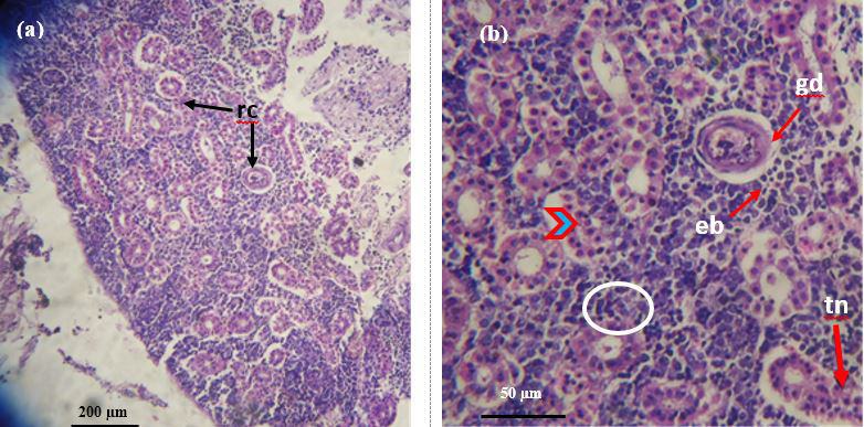
Figure 9:
Photomicrographs of the infested kidney of catla by Argulus japonicus. a) Showing renal corpuscles (rc) (10×); b) Glomerular distension (gd) accompanied with extended bowmans capsule (eb), vacuolation (arrow head) tubular necrosis (tn), increase in the presence of leukocyte cells (circle) (40×).