
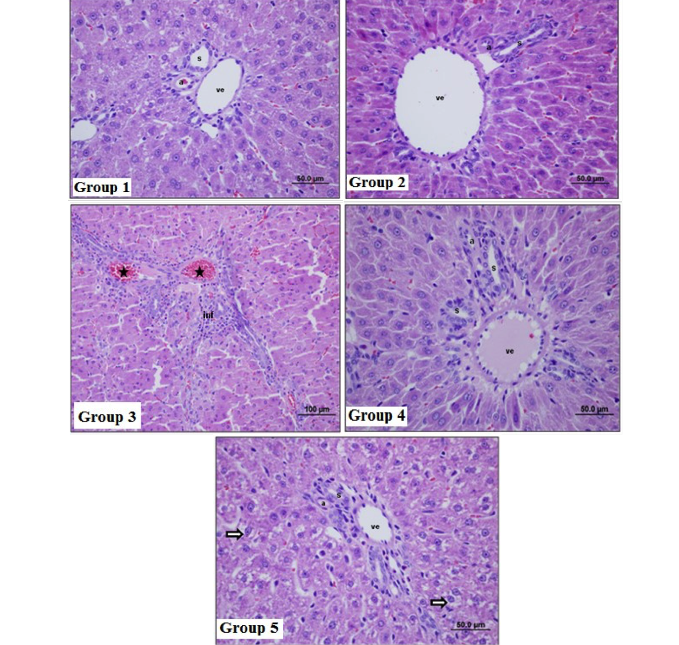
Fig 1
Histological structure of rat liver. Group 1, the control, saline treatment; Group 2, 0.2% DMSO treated liver tissues; Group 3, tissues treated with GalN; Group 4, EA administered liver tissues before GalN treatment; Group 5, tissues administered with GalN before EA. (inf, inflammatory; *, vascular congestion; →, cellular damage; ve, venul; a, arteriole; s, bile duct 50 µm (x40) HE, haematoxylin and eosin stained rat liver tissues.