
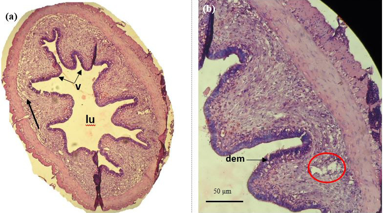
Figure 11:
Histological analysis of mid gut of catla infested with Argulus japonicus. a) Transvers section showing short and stout villi (v) large lumen space (lu) with lamina propria (lp) disruption of mucosal and sub-mucosal tissues (arrow) (10×) b) Damage of enterocytes and microvilli (dem) with large vacuolar space (circle) (40×).