
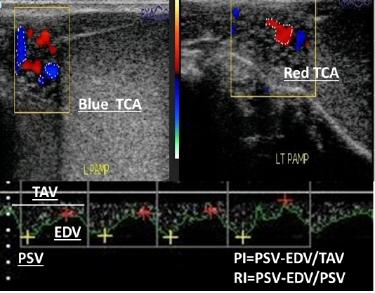
Figure 1
Color mode ultrasonograms showed the measurement of both blue and red testicular colored areas (TCA) either away or toward the probe with the presence of spectral wave graph that determined how to measure both Doppler indices (PI; pulsatility index and RI; resistive index) through different automatic equations using peak systolic velocity (PSV; cm/sec), end diastolic velocity (EDV) and time average to complete the cardiac cycle (TAV; cm/sec).