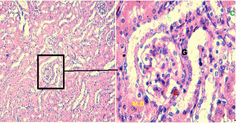
Fig 6
Histological structure of kidney of positive control mouse. Thich black arrows in (x100) showing epithelial crescent squashing the glomerular tufts from all sides, yellow arrow showing necrocitising glomerulus, black arrow in (x400) showing segmental glomerular sclerosis.