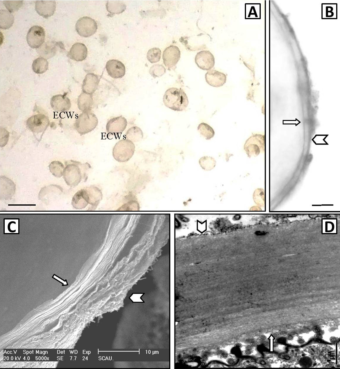
Fig. 1.
Cyst wall of Cryptocaryon irritans viewed by an optical microscope (A, B), SEM (C), and TEM (D). A, a holistic view of empty cyst walls; B, partial view of empty cyst walls; C, cyst wall stripped off in SEM sample preparation, showing the multi-layer structure of the thick cyst wall; D, sectional view of the cyst wall. ECWs represent empty cyst wall. Arrows indicate the inner surface of cyst wall, and Arrowheads indicate the outer surface of the cyst wall. Scale bars = 500 μm (A), 10 μm (B), 10 μm (C), and 1 μm (D).