Figure 2:
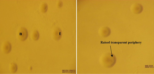
Colonies of Mycoplasma bovis from lung sample on PPLO agar showing ‘fried egg’ colonies with nipples ‘n’ and raised transparent periphery ‘t’ observed under Stereomicroscope (X40).
Colonies of Mycoplasma bovis from lung sample on PPLO agar showing ‘fried egg’ colonies with nipples ‘n’ and raised transparent periphery ‘t’ observed under Stereomicroscope (X40).