
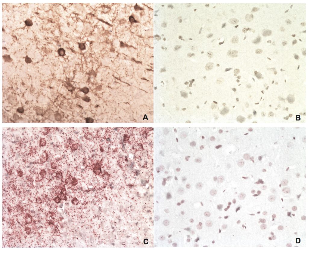
Figure 2
BDV P IHC and in situ PLA on brain tissues from an experimentally infected rat and an uninfected control
A) IHC of BDV P on brain tissue from an experimentally infected rat using a rabbit polyclonal anti-BDV P antibody. The brown staining is BDV P. B) IHC of BDV P on brain tissue from an uninfected control rat. C) In situ PLA of BDV P on brain tissue from the same rat as in A, using rabbit polyclonal and mouse monoclonal anti-BDV P antibodies. The red staining is BDV P. D) In situ PLA of BDV P on brain tissue from the same uninfected control rat as in B. Magnifications: lens x20.