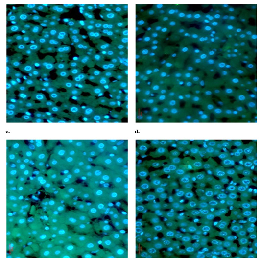
Figure 7
The immunohistochemical staining of CYP-3A4 in rat liver sections was implemented by FITC-conjugated IgG and CYP-3A4 antibody (green) and the nuclei were counterstained with DAPI (blue). Control group (a) and CSO-treated group (b) showed low concentration of CYP-3A4. Group treated with liposomal-doxorubicin (c) showed highly induced CYP-3A4 concentration. CSO/liposomal-doxorubicin group (d) showed highly suppressed CYP-3A4 compared to DOX-treated group. The sections were analyzed under fluorescence microscope (Magnification x400).