
Figure 2
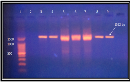
Shows gel electrophoresis, D.N.A. marker: 1 lane, the negative control: 2 lanes, PCR amplification of flic H7 gene: 3-9 lanes, stained with E.B. at 80 volts for 60 minutes, under U.V. light.
Shows gel electrophoresis, D.N.A. marker: 1 lane, the negative control: 2 lanes, PCR amplification of flic H7 gene: 3-9 lanes, stained with E.B. at 80 volts for 60 minutes, under U.V. light.