
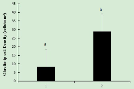
Fig 3
Histogram showing the densities of ghrelin-immunoposive cells (cells/mm2) in the 334 day-old African ostrich pancreas. The number of ghrelin-immunopositive cells were a steady decrease from pancreatic islet to pancreatic acinus. Ghrelin-immunoposive were present throughout the pancreas. a-b, Different letters within the same column indicate significant differences among segments according to Duncan’s multiple range (P≤0.05). 1, pancreatic acinar; 2, islet.