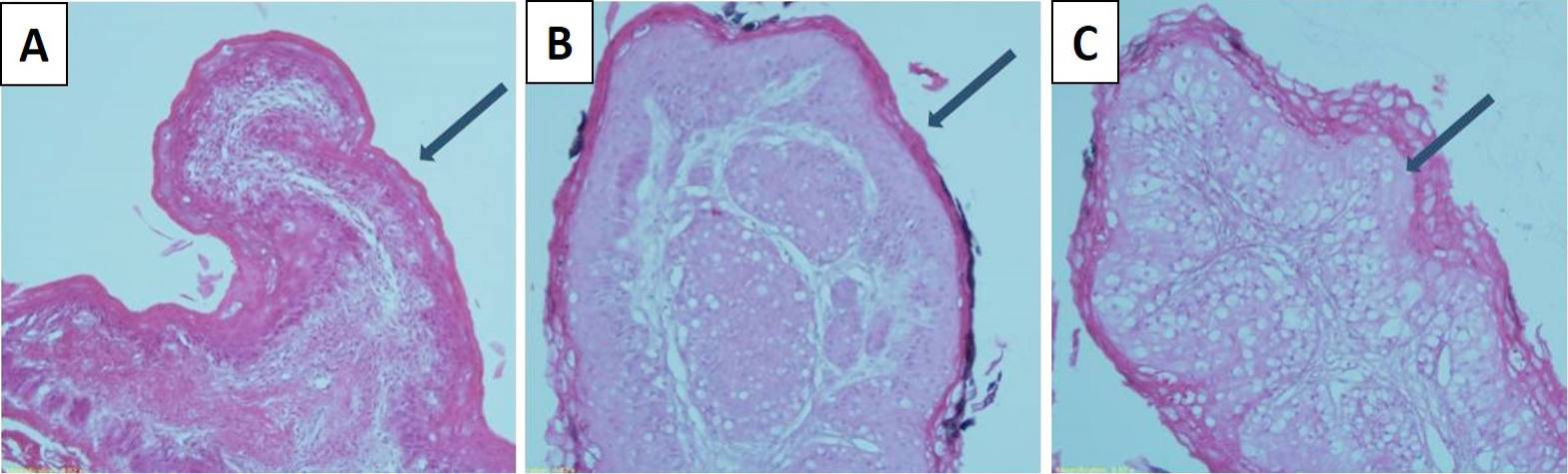
Fig. 2.
Rumen papillae from ventral sacs, arrows show keratin layer of papillae in A, DM; B, CF and C, CFR.
The keratin layer of DM group is very thin, CF group was thick and compact and CFR group was thick and had vacuolated cells (× 20; bar = 500 µm).