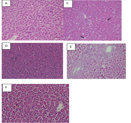
Figure 8
Histological observation of liver tissue. A: Liver tissue in the control group had normal histological appearance. C & D: Liver tissues in lead acetate groups showed that hepatocytes were severely swollen, and have granule denaturation and steatosis. E & F: Cell degeneration of liver tissue becomes mild and improvement was observed in the liver tissues.