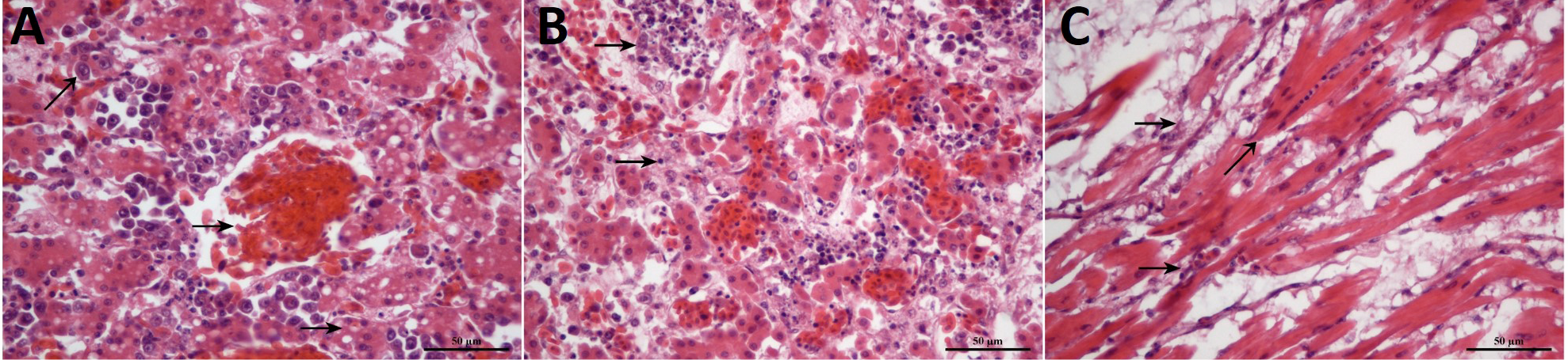
Fig 3
The pathological observation of the congestion in the portal vein with focal lymphoid cells in the liver (A), the degenerative necrosis, hypertrophy with edema on the walls of follicular blood vessels and cells infiltration in the spleen (B) and mild myocardial necrosis, lymphocytic myocarditis, hyperplasia of myocyte nuclei, myocardial infiltration with rupture and myocarditis in the heart (C). Stain, H&E; Magnification, 400x.