
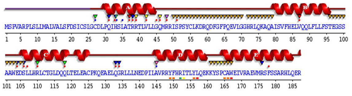
Fig. 5.
Sequence annotations for C. dromedarius IFNα showing the location of α-helices and residues contacting ligand and ions. Secondary structure by homology (), active site residues from PDB site record (▼); residues with contact to ligand (*) and to ions (*).