
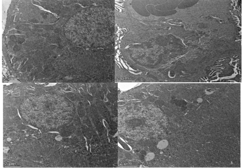
Figure 7
Electron micrograph of a proximal tubule for kidney sections from A: control group (normal ultrastructure of cells) B: adenine group (Large irregular cytosomes contain large numerous myeloid bodies, mitochondria and cisternae of rough edoplasmic reticulum are swollen. Large vacuoles are present near the base of the cell) C: garcinia cambogia D: erlotinib group (both groups show the cell ultra structure is combarable to that of the control).