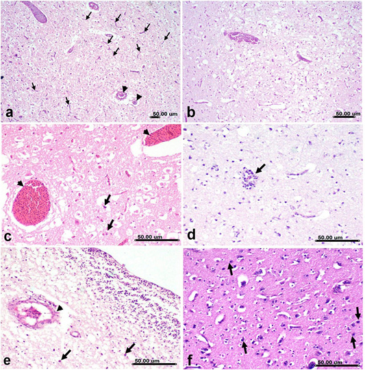
Figure 8
Histological brain sections from H5N8 infected duck showing; a) Congestion and micro- thrombosis of cerebral blood vessels (arrowhead) associated with capillary endothelial proliferation (arrow). b) Spongiosis and vacuolization of cerebral neuropil associate with diffuse gliosis. c) Marked congestion (arrowhead) associated with necrosis of pyramidal neurons involving the cerebellar nucleus(arrow). d) Vasculitis and perivascular lymphocytic cuffing (arrow) associated with gliosis. e) Microthrombosis (arrowhead) and hemorrhage in hippocampus associated with neuronal necrosis (arrow). f) Neuronal degeneration with neuronophagia associated with glisosis of cerebral grey matter. (Stain, H&E)