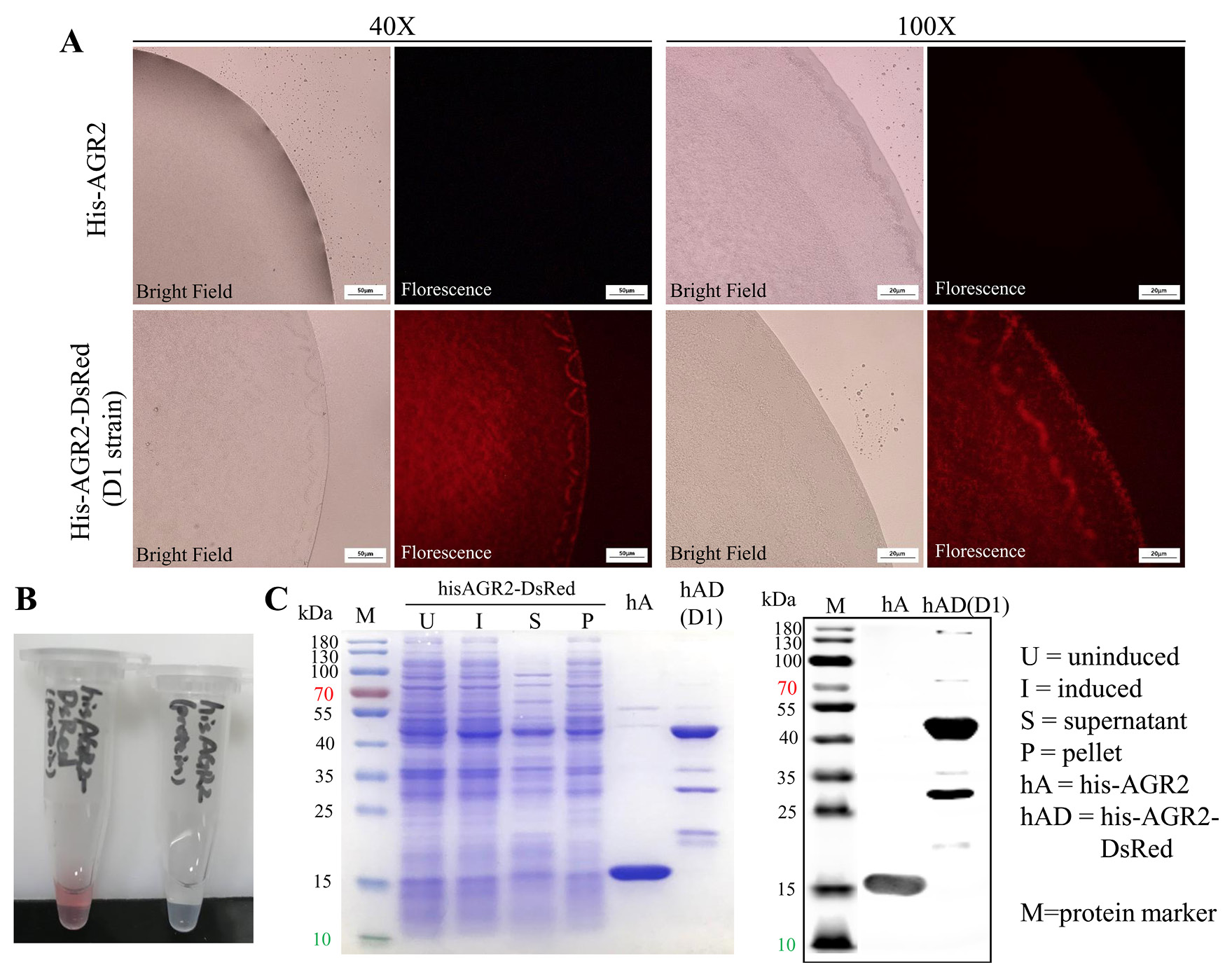
Fig. 6.
Recombinant hAD characterized by fluorescence microscopy and Western blot. (A) Bright field and red fluorescence of bacteria culture of his-AGR2 expression strain and hAD expression D1 strain in both 40× and 100× magnifications. Scale bar, 50μm for 40×magnification and 50μm for 100×magnification. (B) Purified his-AGR2 (up) and hAD (down) protein solution, representing different colors. (C) SDS-PAGE and Western blot result of purified hAD, compared with his-AGR2, along with its original expression in bacteria samples. U, uninduced; I, induced; S, supernatant; P, pellet; hA, his-AGR2; hAD, his-AGR2-DsRed; M, protein marker.