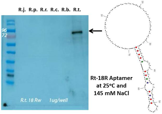
Figure 1
Left panel – aptamer Western blot using the Rt-18R aptamer against protein extracts and R. typhi whole cell lysate all at ~ 1 µg/well. From left to right; molecular weight markers, protein extracts from R. typhi, R. parkeri, R. rickettsii, R. conorii, R. bellii, and an R. typhi whole cell lysate. Right panel – Secondary stem-loop structure of the Rt-18R aptamer determined by energy minimization using UNAFold software with 25oC, 137 mM NaCl, and DNA parameters