
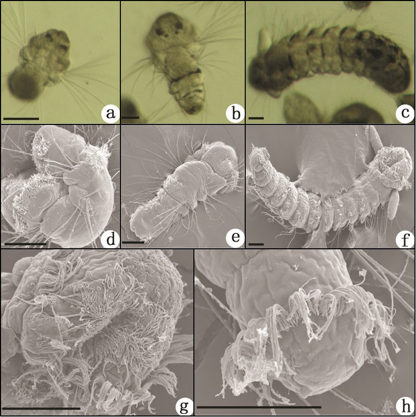
Fig 2
Larvae of Polydora websteri. a–c, Light photographs; d–h, SEM images: a, Three-chaetiger larva, dorsal view, showing two pigmented eyespots on the dorsal surface of the head; b, Five-chaetiger larva, dorsal view, two rows of pigment scattered across the dorsal surface of each chaetiger; c, Thirteen-chaetiger larva, dorsal view; d, Three-chaetiger larva, lateral view, serrated chaetae present on each side of the chaetigers; e, Five-chaetiger larva, dorsal view, the prototroch with a band of fine cilia, encircling the head, with the exception of the dorsal part; f, Thirteen-chaetiger larva, ventral view, a pair of round palps present on both sides of the head; g, Ventral view of the head, showing the vestibule with bundles of cilia; h, Dorsal view of posterior end, the telotroch with a circle of cilia. Scale bars = 50 µm.