
Figure 8
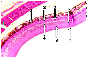
Micrographic of eye in duck shows:1-Retinal pigment epithelium, 2-Photoreceptors and glial cells (Rod and cone cells), 3- Outer nuclear layer, 4-Outer plexiform layer, 5-Inner nuclear layer, 6-Inner plexiform layer, and 7-Ganglion cell layer (H&E stain 400X).