
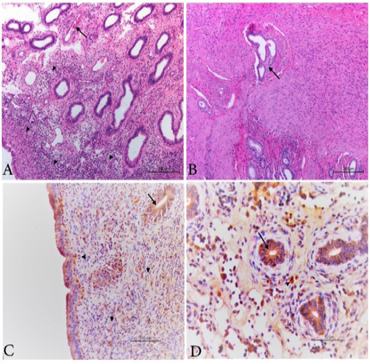
Figure 5
Photomicrograph from uterus of PI animal showing, A) endometritis with submucosal and interglandular infiltration with inflammatory cells (arrow head) as well as congestion of the vasculature (arrow). H&E Bar.200µm. B) Islets from endometrial glands between bundles of uterine muscles (endometriosis) (Arrow). H&E Bar.200µm. C) positive BVDV antigen in the cytoplasm of the glandular epithelium (arrow) and in macrophages at sub epithelial, inter -glandular tissue and in the endometrial glands (arrow head). IHC labelled. Bar.100µm. D) positive brown granules in the cytoplasm of the glandular epithelium (arrow) and in macrophages at the inter -glandular tissue (arrow head). IHC labelled. Bar.50µm.