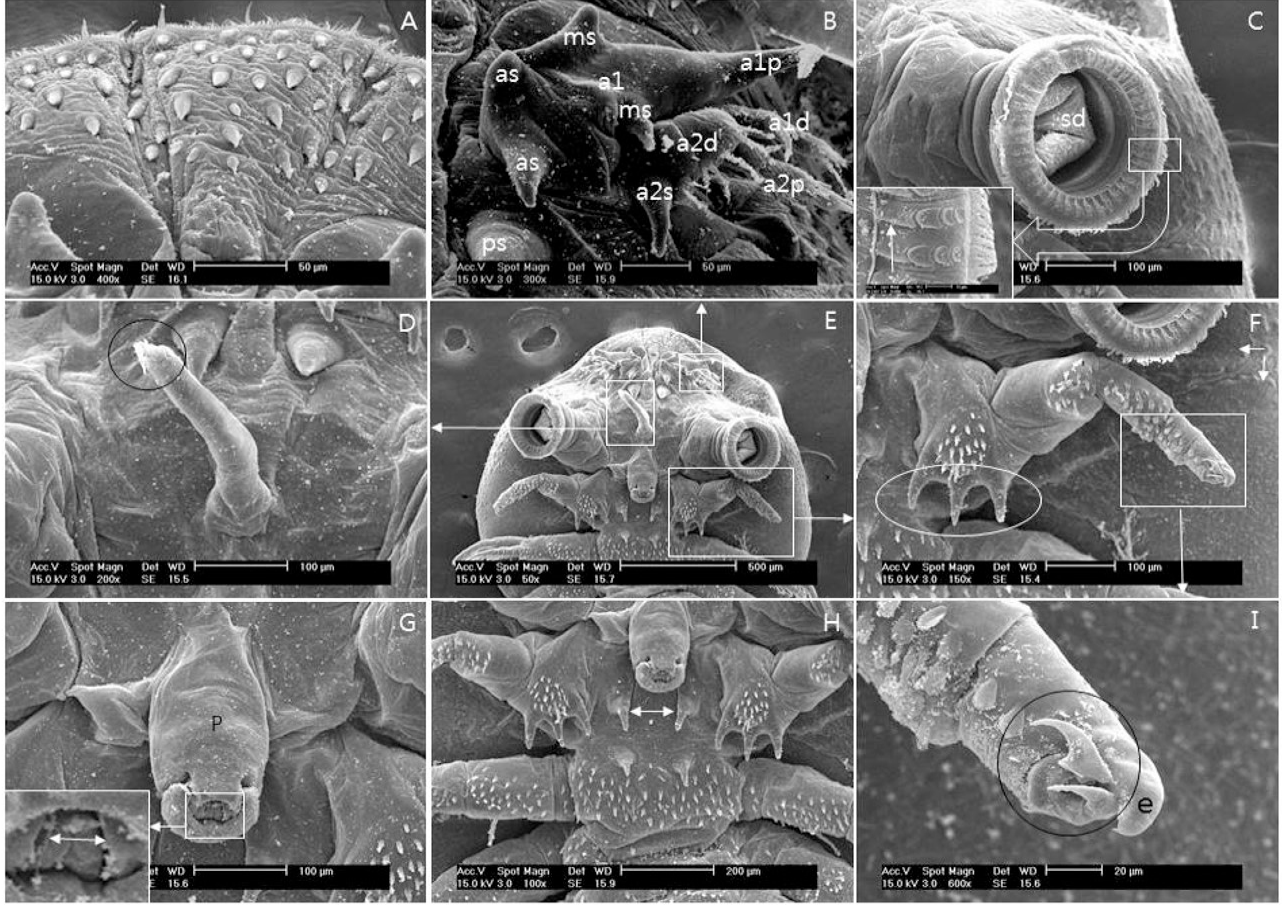
Fig 2
Scanning electron micrographs of anterio-ventral view of male A. japonicus. A, Dentate scales that face towards posterior that occur along the edge of the ventral carapace; B, Antennule and antennae; C, First maxilla. Rim of sucker with supporting rods and sclerites. Basis elongated plate (arrow in square); D, Pre-oral spine, Note the terminal (circle); E, Anterio- ventral part; F, Maxilla with five segments, basal podomere (plate) with three tooth (circle). Two lobes of respiratory area (arrows); G, Proboscis without any dorsal scales. Mouse tube (arrow in square); H, Basement of maxillae and first leg. Basal plate scales of maxillae (arrow); I, Terminal segment of maxilla in a blunt fused extension has two recurved sharp claws (Circle). e, extension; a, antenna; as, anterior spine; a1, first antenna; a1d, first antenna distal part; a2d, second antenna distal part; a2p, second antenna proximal part; a2s, spine of second antenna; ms, median spine; p, proboscis; ps posterior spine; a1p, first antenna proximal hook; sd, suckiong disc.