
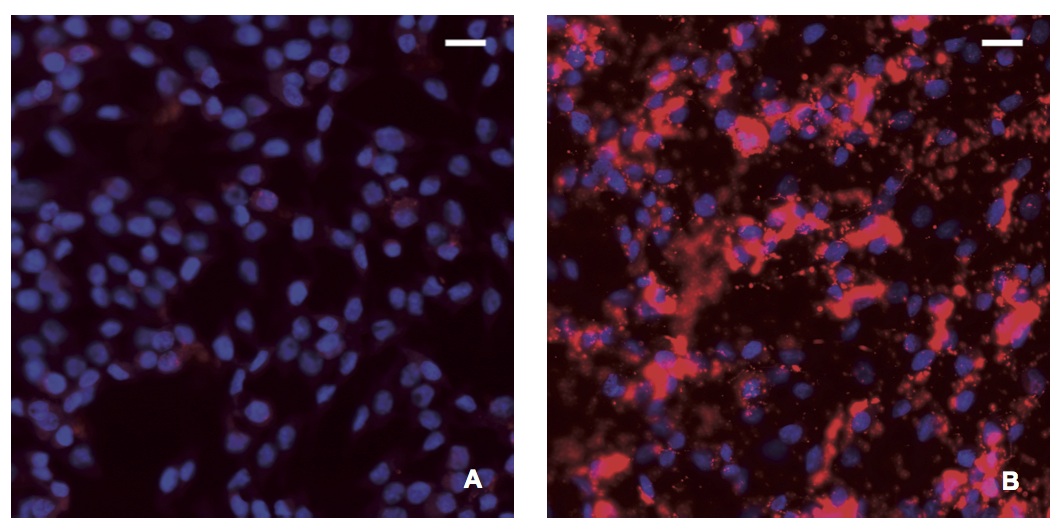
Figure 1
Detection of BDV P using in situ PLA in cell cultures
In situ PLA using a rabbit polyclonal anti-BDV P antibody and a monoclonal anti-BDV P antibody (dilution 1:100) was performed in A) C6 and B) C6BV cell cultures. The in situ PLA signals (BDV P) are seen as red dots, nuclei are blue. Notice the oversaturated staining of BDV P, especially in nuclei of C6BV, resulting from several in situ PLA signals coalescing. Magnifications: lens x20.