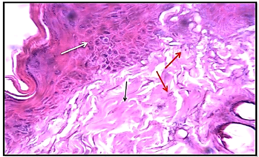
Figure 6
Histological section of of 7th day healing in wound treated group (locally, orally) showed the slight inflammatory cells infiltration (red arrow), mild fibrosis (collagen fibers) (black arrow) and significant keratocytes proliferation (white arrow). (Hematoxylin and Eosin, 10 X).