
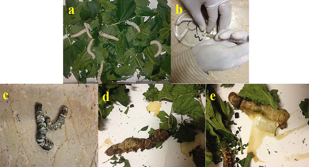
Figure 1
(a) Healthy and uninfected larva of B. mori. (b) Collecting of Haemolymph from caudal horn of B. mori. (c) Infected larva of 2nd day with light brown colour. (d) Larval color changed to coffee brown on 3rd day. (e) Yellowish body fluid discharged from the midgut of B. mori by rupturing the integument after highly infected with B. thuringiensis.