
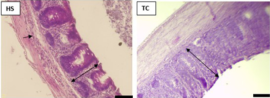
Figure 1
The histological observations in ceca of chickens shown by HE stains. HE-stained specimens were observed under light microscopy. The arrow indicates inflammatory cells infiltrate to mucosa and submucosa. The double-headed arrows indicate the mucosal length. Bar scale is 50 µm.