
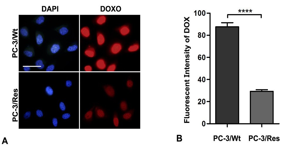
Fig. 2.
Functional analysis of MRP-1 over expression in PC-3/Res cells. (A) Efflux of doxorubicin in PC-3/Wt and PC-3/Res cells monitored using Olympus BX-51 fluorescent microscope. Cells were incubated with 5μM doxorubicin for 2 h and then allowed to efflux for 1h in drug free media. Bar, 50µm. (B) The intracellular fluorescent intensity of doxorubicin post efflux was measured and expressed as mean fluorescent intensity. Error bars represent standard error of mean, n=70; ****P ˂ 0.0001.