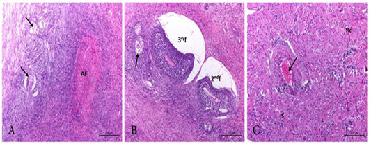
Figure 2
Photomicrograph from ovary of PI cow showing. A) Disrupted primordial follicles (arrow) and obliterative atretic follicle. (AF). H&E. Bar.200µm. B) disrupted secondary and tertiary follicles (2ndf, 3rf). Minute focal granulosa cell tumor (arrow) could be observed. H&E. Bar.200 µm. C) Active corpus luteum with large (L), small(S) regressing cells (Rc). The blood vessel showing congestion and degenerated wall (BVs) (arrow).H&E. Bar.200 µm.