
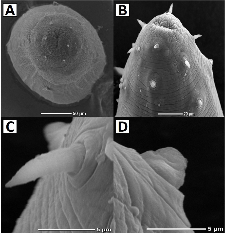
Fig. 4.
Scanning electron micrographs of L4 Eustrongylides tubifex: A, en face view showing slit-like mouth (arrow) and number of papillae; B, anterior end showing arrangement of papillae; C, inner papilla showing narrow base and spine-like apex; D, outer papilla having wide base and nipple-like apex.