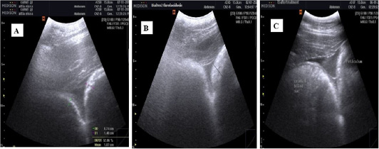
Figure 4
Ultrasonographic images of the camel reticulum visualized from the right paramedian region just behind the sternal pad using 3.5 MHz convex probe (transverse). (A) Before induction of acidosis: The wall of the dorsal ruminal sac wall appeared echogenic line and the reticulum appeared as a half-moon-shaped echogenic structure with an even contour and the contents of dorsal ruminal sac and reticulum appeared hypoechoic and homogenous. (B) After induction of acidosis: The reticulum wall thickness increased. (C) After treatment with Rumitone: The reticular wall thickness decreased compared to pre-acidosis.