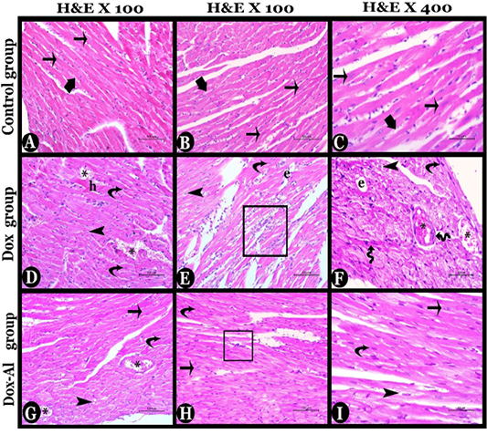
Photomicrographs of histolopathological sections of the heart tissues of all studied mice stained with H&E stain, 1st, and 2nd columns X100, and 3rd column X400: A, B & C) Control group showing normal cardiac architecture, normal arrangement of cylindrical branched myocytes (thin arrows) contained acidophilic cytoplasm and oval central vesicular nuclei. Note, narrow slits (thick arrows) between the myocytes contained fine connective tissue and normal blood vessels. D, E & F) Doxorubicin group showing degenerated condensed myocytes with pyknotic nuclei (curved arrows), deteriorated myocytes with vacuolated cytoplasm infiltrated by inflammatory cells (square), interfibrillar vacuolations (arrowheads), Hyalinosis (h), in addition to damaged blood vessels with congestion (*), proliferating fibroblast (zigzagged arrows) and interstitial edema (e). G, H & I) Almond oil-treated group showing mild degenerative changes appeared in form of few degenerated myocytes with pyknotic nuclei (curved arrows), interfibrillar vacuolations (arrowheads), few inflammatory cells infiltration (square) and vascular degeneration with congestion (*), while most of myocytes appeared as normal (thin arrows).