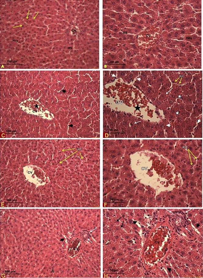
Effects of cadmium and quercetin on the histological sections of male Sprague-Dawley rate. A, B, control rates livers at 10x and 20x magnification, respectfully. A shows the central vein (CV), normal arrangement of hepatocytes (Hcy) along the hepatic cord (arrows) with normal blood sinusoids (BS) and B shows the central vein (CV), normal arrangement of hepatocytes (Hcy) along the hepatic cord (arrows) with normal blood sinusoids (BS); C, D, liver of rates oraly administrated with CdCl2 at 5mg/kg bw/d at 20x (C) and 40x (D) magnification. C shows disorganized hepatic architecture, dilated blood sinusoids (arrow heads), and dilated central vein (CV) (star); D shows dilated central vein (CV) (black star), dilated blood sinusoids (Arrow heads) congested with blood cells. Notice red blood cells (black arrow), Kupffer cells (KC) (yellow arrows), the disintegration in the tissue (white star), and degenerated hepatocytes and necrosis (arrows). E, F, liver of rates oraly administrated with quercetin at 5mg/kg bw/d at 20x (E) and 40x (F) magnification. E shows mild restoration of hepatic tissue. Notice Kupffer cell (KC) (yellow arrows), less disintegration in the central vein (CV) and more organized hepatocytes (star); F shows mild restoration of hepatic tissue. Notice red blood cells (black arrow), Kupffer cells (KC) (yellow arrows), less disintegration in the central vein (CV) and more organized hepatosytes. G, H, liver of rates oraly administrated with quercetin at 70mg/kg bw/d at 20x (G) and 40x (H) magnification. G shows disorganized hepatic architecture with disintegrated and highly congested central vein (CV) (star), inflammatory cell infiltration (white arrows), H shows dilated blood sinusoids (arrow heads) and increase in number of Kupffer cells (yellow arrows); B shows dilated blood sinusoids (arrow heads), necrosis (black arrows) and disintegration and congestion in the central vein (CV) (star) and inflammatory cell infiltration (white arrows). Notice the increase in number of Kupffer cells (yellow arrows).
Magnification: A, 10x; B, C, E, G, 20x; D,F, H, 40x.
Stain: Haematoxylin and Eosin