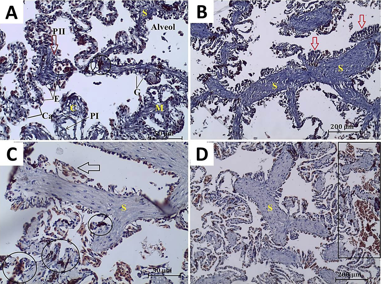
Fig. 2.
A, normal alveolar construction of P. ridibundus collected from DB1. Septum (S), type I and II pneumocytes (PI, PII), erythrocytes (E), goblet cells (G), capillaries (Ca), alveol, melanomacrophage aggregate (encircled), connective (C) and muscle tissue (M). B, C and D, histopathological alterations of the lung of P. ridibundus: B: Samples of DB2; C, D: Samples of SV. B, thickness of alveolar septum and hyperplasia of alveolar epithelium (arrows). C, dilated blood capillaries (arrow) and melanomacrophage aggregation (encircled). D, congestion (squared), (H&E).